

Where Do You Find Chemical And Voltage Channels/gates And What Do These Gates Control?
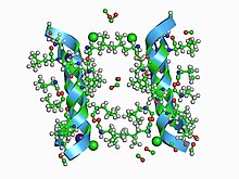
An animated representation of the molecular construction of a simple ion aqueduct
In electrophysiology, the term gating refers to the opening (activation) or closing (by deactivation or inactivation) of ion channels.[one] This change in conformation is a response to changes in transmembrane voltage.[2]
When ion channels are in a 'closed' (non-conducting) state, they are impermeable to ions and do non behave electric current. When ion channels are in their open state, they conduct electrical current by assuasive specific types of ions to pass through them, and thus, beyond the plasma membrane of the cell. Gating is the procedure past which an ion channel transitions between its open and closed states.[3]
A multifariousness of cellular changes can trigger gating, depending on the ion aqueduct, including changes in voltage beyond the cell membrane (voltage-gated ion channels), chemicals interacting with the ion channel (ligand-gated ion channels), changes in temperature,[4] stretching or deformation of the cell membrane, addition of a phosphate group to the ion channel (phosphorylation), and interaction with other molecules in the prison cell (eastward.g., G proteins).[5] The rate at which whatever of these gating processes occurs in response to these triggers are known as the kinetics of gating. Some drugs and many ion channel toxins human action equally 'gating modifiers' of voltage-gated ion channels by changing the kinetics of gating.[six]
The voltage-gated ion channels of the action potential are oft described as having four gating processes: activation, deactivation, inactivation, and reactivation (besides called 'recovery from inactivation'). Activation is the procedure of opening the activation gate, which occurs in response to the voltage inside the cell membrane (the membrane potential) becoming more positive with respect to the outside of the cell (depolarization), and 'deactivation' is the opposite process of the activation gate endmost in response to the inside of the membrane condign more negative (repolarization). 'Inactivation' is the endmost of the inactivation gate, and occurs in response to the voltage inside the membrane becoming more positive, just more than slowly than activation. 'Reactivation' is the opposite of inactivation, and is the procedure of reopening the inactivation gate.[vii]
These voltage-dependent changes in role are critical for a large number of processes in excitable and nonexcitable cells.[two]
Activation [edit]
Voltage-gated ion channels [edit]
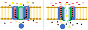
Voltage-gated ion channel. When the membrane is polarized, the voltage sensing domain of the channel shifts, opening the aqueduct to ion flow (ions represented by yellow circles).
Voltage-gated ion channels open and shut in response to the electrical potential beyond the cell membrane. Portions of the aqueduct domain human action as voltage sensors. As the membrane potential changes, this results in changes in electrostatic forces, moving these voltage-sensing domains. This changes the conformation of other elements of the channel to either the open or closed position.[8] When they move from the closed position to the open position, this is called "activation." Voltage-gated ion channels underlie many of the electrical behaviors of the cell, including action potentials, resting membrane potentials, and synaptic transmission.[ix]
Voltage-gated ion channels are often specific to ions, including Na+, Grand+, Ca2+, and Cl−. Each of these ions plays an important role in the electrical behavior of the jail cell.[nine] The gates also accept unique properties with important physiological implications. For case, Na+ channels open up and shut apace, while Yard+ gates open up and close much more slowly. The difference in speed between these channels underlies the depolarization and repolarization phases of the activeness potential.[ten]
Na+ Channels [edit]
Voltage Gated Sodium (Na+) channels are significant when it comes to propagating the action potentials in neurons and other excitable cells, mostly being used for the propagation of action potential in axons, muscle fibers and the neural somatodendritic compartment.[xi] Sodium(Na+) channels are some of the main ion channels responsible for action potentials.[9] Being complex, they are made of bigger α subunits that are and then paired with ii smaller β subunits.[xi] They contain transmembrane segments known as S1-6. The charged S4 segments are the channels voltage sensors. When exposed to a certain minimum potential difference, the S4 segments move across the membrane.[12] This causes movement of the S4-S5 linker, which causes the S5-S6 linker to twist and opens the aqueduct.[13]
M+ Channels [edit]
Potassium (K+) channels play a large part in setting the resting membrane potential.[9] When the cell membrane depolarizes, the intracellular part of the channel becomes positively charged, which causes the channel'southward open up configuration to become a more stable state than the closed configuration. There are a few models of potassium channel activation:
- The sliding helix model posits that the potassium aqueduct opens due to a screwing motion by its S4 helix.
- The paddle model posits that the S3 and S4 helices of the channel form "paddles" that movement through the depolarized membrane and pull the S5 helix away from the channel's opening.
- The transport model posits that a focused electrical field causes charged particles to move across the channel with only a small movement of the S4 helix.
- The model of coordinated movement of helices posits that the S4 and S5 helices both rotate, and the S4-S5 linker causes the S6 helix to movement, opening the channel.
- The consensus model is an boilerplate of the higher up models that helps reconcile them with experimental data.[14]
Ca2+ Channels [edit]
Calcium (Caii+) channels regulate the release of neurotransmitters at synapses, control the shape of action potentials made by sodium channels, and in some neurons, generate action potentials.[9] Calcium channels consist of half dozen transmembrane helices. S4 acts as the voltage sensor by rotating when exposed to sure membrane potentials, thereby opening the channel.[fifteen]

Calcium release causes a strong attraction between multiple proteins including synaptobrevin and SNARE proteins to pull the neurotransmitter vesicle to the membrane and release its contents into the synaptic scissure
Neurotransmitters are initially stored and synthesized in vesicles at the synapse of a neuron. When an action potential occurs in a cell, the electric signal reaches the presynaptic terminal and the depolarization causes calcium channels to open, releasing calcium to travel downwardly its electrochemical gradient. This influx of calcium after is what causes the neurotransmitter vesicles to fuse with the presynaptic membrane.[16] The calcium ions initiate the interaction of obligatory cofactor proteins with SNARE proteins to grade a SNARE circuitous.[16] These SNARE complexes mediate vesicle fusion by pulling the membranes together, leaking the neurotransmitters into the synaptic cleft. The neurotransmitter molecules can then betoken the next cell via receptors on the post synaptic membrane. These receptors tin either act as ion channels or GPCR (G-Protein Coupled Receptors).[17] In general the neurotransmitter can either crusade an excitatory or inhibitory response, depending on what occurs at the receptor.
Cl− Channels
Chloride channels are another grouping of voltage gated ion channels, of which are less understood. They are involved with processes such as skeletal and cardiac smoothen muscle, cell volume regulation, the jail cell bicycle, and apoptosis.[xviii] One major family of chloride proteins are chosen CLC proteins- common channels and transporters for basic physiological processes in mammals. CLC channels act as tedious gated channels; hydrogen ions are exchanged for an influx of chloride ions, assuasive the anions to travel via their electrochemical gradient.[19] The voltage dependent C1C-1 chloride channel is homologous dimer which falls under this family, and is seen predominantly in skeletal muscle fibers.[20] With this channel the correct depolarization and repolarization via chloride ions is essential for propagation of an activeness potential.[18]
Ligand-gated ion channels [edit]
Ligand-gated ion channels are found on postsynaptic neurons. By default, they assume their closed conformation. When the presynaptic neuron releases neurotransmitters at the cease of an activity potential, they bind to ligand-gated ion channels. This causes the channels to presume their open up conformation, allowing ions to flow through the channels down their concentration gradient. Ligand-gated ion channels are responsible for fast synaptic transmission in the nervous system and at the neuromuscular junction.[21] Each ligand-gated ion channel has a wide range of receptors with differing biophysical properties as well as patterns of expression in the nervous system.[22]
Inactivation [edit]
Inactivation is when the flow of ions is blocked by a machinery other than the closing of the channel.[viii] A channel in its open up state may stop allowing ions to flow through, or a channel in its airtight land may exist preemptively inactivated to prevent the flow of ions.[23] Inactivation typically occurs when the cell membrane depolarize, and ends when the resting potential is restored.[8]
In sodium channels, inactivation appears to exist the result of the actions of helices III-Half-dozen, with Iii and IV acting as a sort of hinged lid that block the channel. The verbal machinery is poorly understood, only seems to rely on a particle that has a high affinity for the exposed inside of the open up aqueduct.[24] Rapid inactivation allows the channel to halt the menstruation of sodium very soon subsequently assuming its open up conformation.[25]
Ball and chain inactivation [edit]
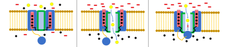
Voltage-gated ion aqueduct in its closed, open, and inactivated states. The inactivated channel is even so in its open state, but the ball domain blocks ion permeation.
The ball and chain model, as well known as Northward-type inactivation or hinged lid inactivation, is a gating machinery for some voltage-gated ion channels. Voltage-gated ion channels are composed of iv[ dubious ] α subunits, i or more than of which will accept a ball domain located on its cytoplasmic N-terminus.[26] The ball domain is electrostatically attracted to the inner channel domain. When the ion aqueduct is activated, the inner channel domain is exposed, and inside milliseconds the chain will fold and the ball will enter the channel, occluding ion permeation.[27] The channel returns to its closed country, blocking the channel domain, and the ball leaves the pore.[28]
Deactivation [edit]

Equally the membrane potential returns to its resting value, the voltage differential is non sufficient to proceed the channel in its open state, causing the channel to shut.
Deactivation is the return of an ion channel to its airtight conformation. For voltage-gated channels this occurs when the voltage differential that originally acquired the channel to open returns to its resting value.[29]
In voltage-gated sodium channels, deactivation is necessary to recover from inactivation.[24]
In voltage gated potassium channels, the opposite is true, and deactivation slows the channel's recovery from activation.[30] The closed conformation is assumed by default, and involves the fractional straightening of helix VI by the Four-V linker. The mechanisms that cause opening and endmost are not fully understand. The closed conformation appears to be a higher energy conformation than the open conformation, which may likewise assistance explain how the ion channel activates.[31]
Quantification [edit]
Gating charge can be calculated past solving Poisson'due south equation. Recent studies have suggested a molecular dynamics simulation-based method to make up one's mind gating charge past measuring electrical capacitor properties of membrane-embedded proteins.[2] Activity of ion channels located in the plasma membrane can be measured by but attaching a glass capillary electrode continuously with the membrane.[32] Other ion channels located in the membranes of mitochondria, lysosomes, and the Golgi appliance can be measured by an emergent technique that involves the use of an bogus bilayer lipid membrane attached to a 16 electrode device that measures electrical action.[32]
Run across too [edit]
- Synaptic gating
- Synaptic potentials
References [edit]
- ^ Alberts, Bruce; Bray, Dennis; Lewis, Julian; Raff, Martin; Roberts, Keith; Watson, James D. (1994). Molecular biology of the prison cell . New York: Garland. pp. 523–547. ISBN978-0-8153-1620-6.
- ^ a b c Machtens, Jan-Philipp; Briones, Rodolfo; Alleva, Claudia; de Groot, Bert L.; Fahlke, Christoph (2017-04-11). "Gating Charge Calculations by Computational Electrophysiology Simulations". Biophysical Periodical. 112 (vii): 1396–1405. Bibcode:2017BpJ...112.1396M. doi:10.1016/j.bpj.2017.02.016. ISSN 0006-3495. PMC5389965. PMID 28402882.
- ^ Goychuk, Igor; Hänggi, Peter (2002-03-nineteen). "Ion aqueduct gating: A beginning-passage time analysis of the Kramers type". Proceedings of the National Academy of Sciences of the United States of America. 99 (6): 3552–3556. arXiv:physics/0111187. Bibcode:2002PNAS...99.3552G. doi:10.1073/pnas.052015699. ISSN 0027-8424. PMC122561. PMID 11891285.
- ^ Cesare P, Moriondo A, Vellani V, McNaughton PA (July 1999). "Ion channels gated by heat". Proc. Natl. Acad. Sci. UsaA. 96 (xiv): 7658–63. Bibcode:1999PNAS...96.7658C. doi:10.1073/pnas.96.14.7658. PMC33597. PMID 10393876.
- ^ Hille, Bertil (2001). Ion channels of excitable membranes. Sunderland, Mass: Sinauer. ISBN978-0-87893-321-1.
- ^ Waszkielewicz, A.M; Gunia, A; Szkaradek, Northward; Słoczyńska, G; Krupińska, Southward; Marona, H (April 2013). "Ion Channels as Drug Targets in Central Nervous Organization Disorders". Current Medicinal Chemistry. 20 (10): 1241–1285. doi:10.2174/0929867311320100005. ISSN 0929-8673. PMC3706965. PMID 23409712.
- ^ Ahern, Christopher A.; Payandeh, Jian; Bosmans, Frank; Chanda, Businesswoman (Jan 2016). "The hitchhiker'south guide to the voltage-gated sodium channel milky way". The Journal of General Physiology. 147 (1): 1–24. doi:x.1085/jgp.201511492. ISSN 0022-1295. PMC4692491. PMID 26712848.
- ^ a b c Bähring, Robert; Covarrubias, Manuel (2011-02-01). "Mechanisms of airtight-state inactivation in voltage-gated ion channels". The Journal of Physiology. 589 (Pt 3): 461–479. doi:ten.1113/jphysiol.2010.191965. ISSN 0022-3751. PMC3055536. PMID 21098008.
- ^ a b c d e Purves, Dale; Augustine, George J.; Fitzpatrick, David; Katz, Lawrence C.; LaMantia, Anthony-Samuel; McNamara, James O.; Williams, Due south. Marker (2001). "Voltage-Gated Ion Channels". Neuroscience. 2nd Edition.
- ^ Grider, Michael H.; Glaubensklee, Carolyn S. (2019), "Physiology, Activity Potential", StatPearls, StatPearls Publishing, PMID 30844170, retrieved 2019-10-29
- ^ a b Mantegazza, Massimo; Catterall, William A. (2012), Noebels, Jeffrey 50.; Avoli, Massimo; Rogawski, Michael A.; Olsen, Richard W. (eds.), "Voltage-Gated Na+ Channels: Structure, Office, and Pathophysiology", Jasper's Basic Mechanisms of the Epilepsies (fourth ed.), National Center for Biotechnology Information (U.s.a.), PMID 22787615, retrieved 2019-11-03
- ^ Sula, Altin; Booker, Jennifer; Ng, Leo C. T.; Naylor, Claire Due east.; DeCaen, Paul Thousand.; Wallace, B. A. (2017-02-16). "The complete structure of an activated open sodium aqueduct". Nature Communications. 8 (1): 14205. Bibcode:2017NatCo...814205S. doi:10.1038/ncomms14205. ISSN 2041-1723. PMC5316852. PMID 28205548.
- ^ Catterall, William A. (2013-11-14). "Structure and function of voltage-gated sodium channels at atomic resolution". Experimental Physiology. 99 (one): 35–51. doi:10.1113/expphysiol.2013.071969. ISSN 0958-0670. PMC3885250. PMID 24097157.
- ^ Grizel, A. 5.; Glukhov, Thousand. Due south.; Sokolova, O. S. (Oct–Dec 2014). "Mechanisms of Activation of Voltage-Gated Potassium Channels". Acta Naturae. 6 (4): 10–26. doi:x.32607/20758251-2014-6-4-10-26. PMC4273088. PMID 25558391.
- ^ Catterall, William A. (August 2011). "Voltage-Gated Calcium Channels". Cold Spring Harbor Perspectives in Biological science. 3 (8): a003947. doi:x.1101/cshperspect.a003947. ISSN 1943-0264. PMC3140680. PMID 21746798.
- ^ a b Südhof, Thomas C. (January 2012). "Calcium Command of Neurotransmitter Release". Cold Jump Harbor Perspectives in Biology. 4 (1): a011353. doi:ten.1101/cshperspect.a011353. ISSN 1943-0264. PMC3249630. PMID 22068972.
- ^ Yoon, Tae-Young; Lu, Xiaobing; Diao, Jiajie; Lee, Soo-Min; Ha, Taekjip; Shin, Yeon-Kyun (June 2008). "Complexin and Ca 2+ stimulate SNARE-mediated membrane fusion". Nature Structural & Molecular Biology. fifteen (vii): 707–713. doi:ten.1038/nsmb.1446. ISSN 1545-9985. PMC2493294. PMID 18552825.
- ^ a b "Chloride channels". British Periodical of Pharmacology. 158 (Suppl 1): S130–S134. November 2009. doi:10.1111/j.1476-5381.2009.00503_6.x. ISSN 0007-1188. PMC2884561.
- ^ Accardi, Alessio; Picollo, Alessandra (Baronial 2010). "CLC channels and transporters: proteins with borderline personalities". Biochimica et Biophysica Acta (BBA) - Biomembranes. 1798 (8): 1457–1464. doi:ten.1016/j.bbamem.2010.02.022. ISSN 0006-3002. PMC2885512. PMID 20188062.
- ^ Imbrici, Paola; Altamura, Concetta; Pessia, Mauro; Mantegazza, Renato; Desaphy, Jean-François; Camerino, Diana Conte (2015-04-27). "ClC-i chloride channels: state-of-the-art inquiry and futurity challenges". Frontiers in Cellular Neuroscience. 9: 156. doi:10.3389/fncel.2015.00156. ISSN 1662-5102. PMC4410605. PMID 25964741.
- ^ Alexander, SPH; Mathie, A; Peters, JA (November 2011). "Ligand-Gated Ion Channels". British Periodical of Pharmacology. 164 (Suppl 1): S115–S135. doi:10.1111/j.1476-5381.2011.01649_4.ten. ISSN 0007-1188. PMC3315629.
- ^ Alexander, SPH; Mathie, A; Peters, JA (2011). "Ligand-Gated Ion Channels". Br J Pharmacol. 164 (Suppl 1): S115–S135. doi:x.1111/j.1476-5381.2011.01649_4.x. PMC3315629.
- ^ Armstrong, Clay M. (2006-11-21). "Na channel inactivation from open up and closed states". Proceedings of the National University of Sciences. 103 (47): 17991–17996. Bibcode:2006PNAS..10317991A. doi:ten.1073/pnas.0607603103. ISSN 0027-8424. PMC1693860. PMID 17101981.
- ^ a b Kuo, Chung-Mentum; Edible bean, Bruce P. (1994-04-01). "Na+ channels must deactivate to recover from inactivation". Neuron. 12 (4): 819–829. doi:ten.1016/0896-6273(94)90335-ii. ISSN 0896-6273. PMID 8161454. S2CID 41285799.
- ^ Yu, Frank H; Catterall, William A (2003). "Overview of the voltage-gated sodium channel family". Genome Biological science. iv (3): 207. doi:10.1186/gb-2003-iv-3-207. ISSN 1465-6906. PMC153452. PMID 12620097.
- ^ "Modulation of One thousand+ aqueduct North-type inactivation by sulfhydration through hydrogen sulfide and polysulfides". rdcu.exist . Retrieved 2018-11-22 .
- ^ Holmgren, M.; Jurman, Thou. East.; Yellen, G. (September 1996). "N-blazon inactivation and the S4-S5 region of the Shaker M+ channel". The Journal of General Physiology. 108 (three): 195–206. doi:ten.1085/jgp.108.3.195. ISSN 0022-1295. PMC2229322. PMID 8882863.
- ^ Bénitah, J. P.; Chen, Z.; Balser, J. R.; Tomaselli, G. F.; Marbán, Eastward. (1999-03-01). "Molecular dynamics of the sodium channel pore vary with gating: interactions between P-segment motions and inactivation". The Periodical of Neuroscience. nineteen (v): 1577–1585. doi:10.1523/JNEUROSCI.xix-05-01577.1999. ISSN 0270-6474. PMC6782169. PMID 10024345.
- ^ Bähring, Robert; Covarrubias, Manuel (2011-01-28). "Mechanisms of closed-country inactivation in voltage-gated ion channels". The Periodical of Physiology. 589 (3): 461–479. doi:x.1113/jphysiol.2010.191965. ISSN 0022-3751. PMC3055536. PMID 21098008.
- ^ Kuo, Chung-Mentum (1997-05-fifteen). "Deactivation Retards Recovery from Inactivation in Shaker 1000+ Channels". The Journal of Neuroscience. 17 (ten): 3436–3444. doi:ten.1523/JNEUROSCI.17-10-03436.1997. ISSN 0270-6474. PMC6573675. PMID 9133369.
- ^ Fowler, Philip Due west.; Sansom, Mark Due south. P. (2013-05-21). "The pore of voltage-gated potassium ion channels is strained when closed". Nature Communications. 4 (1): 1872. Bibcode:2013NatCo...four.1872F. doi:10.1038/ncomms2858. ISSN 2041-1723. PMC3674235. PMID 23695666.
- ^ a b Kamiya, Koki; Osaki, Toshihisa; Nakao, Kenji; Kawano, Ryuji; Fujii, Satoshi; Misawa, Nobuo; Hayakawa, Masatoshi; Takeuchi, Shoji (2018-11-30). "Electrophysiological measurement of ion channels on plasma/organelle membranes using an on-chip lipid bilayer system". Scientific Reports. 8 (i): 17498. Bibcode:2018NatSR...817498K. doi:10.1038/s41598-018-35316-4. ISSN 2045-2322. PMC6269590. PMID 30504856.

Where Do You Find Chemical And Voltage Channels/gates And What Do These Gates Control?,
Source: https://en.wikipedia.org/wiki/Gating_%28electrophysiology%29
Posted by: woodscoubjecruir1961.blogspot.com
0 Response to "Where Do You Find Chemical And Voltage Channels/gates And What Do These Gates Control?"
Post a Comment